Contributing to improvements in diagnostic accuracy by promoting the use of CR that’s effective in the clinical practice of bovine medicine
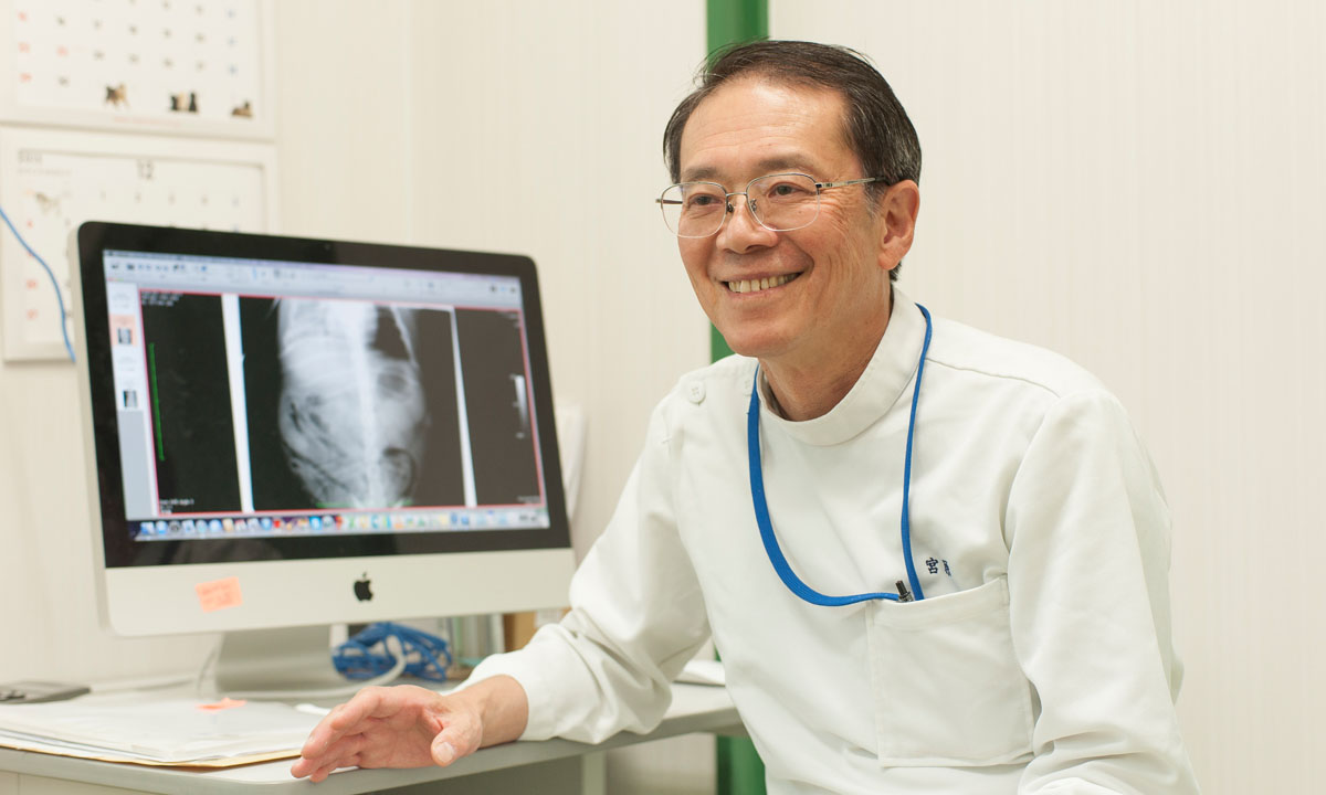
Professor Kazuro MIYAHARA
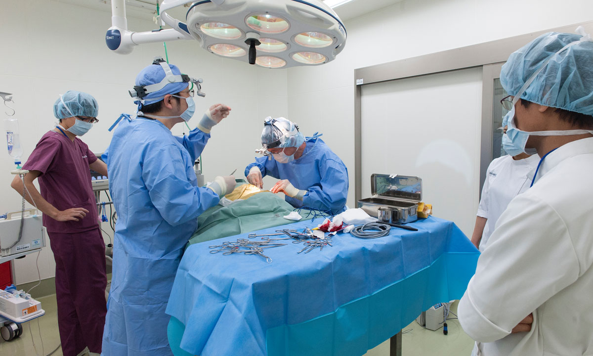
Professor Miyahara has been with the university for more than 40 years, including his days as an undergraduate student. During those years, the transition from analog to digital has occurred. Major changes have also occurred in medical practice for industrial animals, which is his field of research. One such change is the introduction of X-ray fluoroscopy, which has enabled visual diagnoses using images in place of the conventional method of depending on sounds heard through stethoscopes. “The university introduced an image diagnosis vehicle for the X-ray fluoroscopy of industrial animals in 1980,” the professor says. “It was the only one of its kind in the world, and it was only thanks to advanced Japanese technology that is could be made. The advantages of fluoroscopy include less burden placed on cows and the avoidance of unnecessary examinations.” Professor Miyahara talks about the advantages of the test method. “Barium must be used when testing humans, but no contrast agent is necessary for cows, as the contents of the rumen, which is the largest of the four compartments of the cow’s stomach, serve as the background and the other parts of the digestive tract are tucked to one side.” And with equipment for ultrasonography, X-ray photography, phonocardiography, electrocardiography, endoscopies and other tests, the vehicle is used not only for the health examinations of cows, but also for the important post-graduate education of veterinarians. Although they can’t use the vehicle every day to diagnose different kinds of diseases, experience in the vehicle helps them associate sounds heard through stethoscopes and the feel of the rectum with visual images. Professor Miyahara says, “After such experience, they become able to imagine the inside of the body whenever they examine a cow. It’s a true example of a picture being worth a thousand words.” A second diagnostic vehicle, introduced in 1996, is dispatched mainly to eastern Hokkaido for three or four days a month. The vehicle has also been used for training at the request of a veterinary college.
Professor Miyahara has been placing special emphasis on expanding the introduction of a digital x-ray system called computed radiography (CR). Together with the manufacturer, he’s been engaged in developing both hardware and software for this system, which is also installed in the diagnostic vehicle. “Instead of film, the CR system uses an imaging plate that’s irradiated with X-rays to project image information on a monitor. It doesn’t require a developing solution, a fixer or a darkroom.” The professor expresses his expectations for the spread of CR. “A major advantage of CR is that clear images can be obtained even under different radiographic conditions. It’s also possible to diagnose the conditions of bones and soft tissue with a single image. I believe CR will dramatically increase the demand for radiography and contribute to improvements in the accuracy of clinical diagnoses.” Conventional radiographic inspections were often useless, as the images were unclear even though veterinarians went to the trouble of taking the equipment to farms and positioning cows for radiography. Since cows are industrial animals and cost reductions are required, radiography has gradually become unpopular. The CR system promises to be a game-changer.
However, since CR is expensive, only five agricultural mutual aid associations in Hokkaido have introduced it. So Professor Miyahara wrote the book Normal Radiographic Images of Dairy Cows, which presents the limbs of cows of different ages through 156 images from various angles and explanatory text, to support more farmers. The book was co-written with Hiroshi Omura, a graduate of the university. It’s intended to help veterinarians give easy-to-understand explanations to farmers while they compare their own radiographic images to the photos in the book. Along with CR, it will certainly be way of improving diagnostic accuracy in bovine medicine.